Introduction
In the ever-evolving field of medical diagnostics, ultrasonography has emerged as a powerful tool that offers a non-invasive and safe way to visualize the internal structures of the human body. This article delves into the fascinating world of ultrasonography, exploring its principles, applications, benefits, and limitations.
Understanding Ultrasonography
Ultrasonography, commonly known as ultrasound, is a medical imaging technique that uses high-frequency sound waves to create real-time images of the body’s internal structures. Unlike X-rays, which use ionizing radiation, ultrasound is considered safer and is widely used in various medical specialties.
How Does Ultrasonography Work?
Generation of Sound Waves
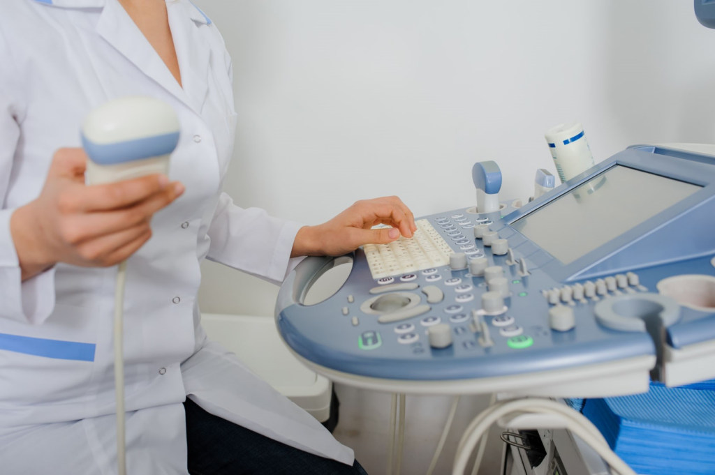
Ultrasonography begins with the generation of sound waves by a transducer. This handheld device emits sound waves that are beyond the range of human hearing.
Reflection and Detection
When these sound waves encounter different tissues and organs within the body, they bounce back (reflect) to the transducer. The transducer detects these echoes and sends the information to a computer, which processes it into images.
Applications of Ultrasonography
Obstetrics and Gynecology
One of the most well-known uses of ultrasonography is in obstetrics, where it allows expectant parents to see images of their developing fetus. It is also crucial for monitoring pregnancies and detecting any potential issues.
Abdominal Imaging
Ultrasonography is widely used to examine abdominal organs such as the liver, kidneys, and gallbladder. It helps in diagnosing conditions like gallstones, liver disease, and kidney abnormalities.
Cardiac Ultrasound
In cardiology, ultrasonography is used for echocardiograms, providing detailed images of the heart’s structure and function. It aids in diagnosing heart diseases and assessing cardiac function.
Advantages of Ultrasonography
Safety
Ultrasonography is non-invasive and does not involve exposure to ionizing radiation, making it safe for all age groups, including pregnant women.
Real-Time Imaging
One of its major advantages is the ability to obtain real-time images, allowing doctors to observe moving structures such as a beating heart or a developing fetus.
Versatility
It can be used for a wide range of medical purposes, from diagnosing diseases to guiding surgical procedures.
Limitations and Considerations
Operator Dependency
The quality of ultrasound images can vary depending on the operator’s skill and experience.
Limited Tissue Penetration
Ultrasonography may not be suitable for imaging structures deep within the body, as sound waves can be blocked or distorted by bone or air.
Conclusion
Ultrasonography has revolutionized the field of medical imaging with its safety, versatility, and real-time imaging capabilities. While it has its limitations, its widespread use in various medical specialties continues to benefit patients worldwide.
